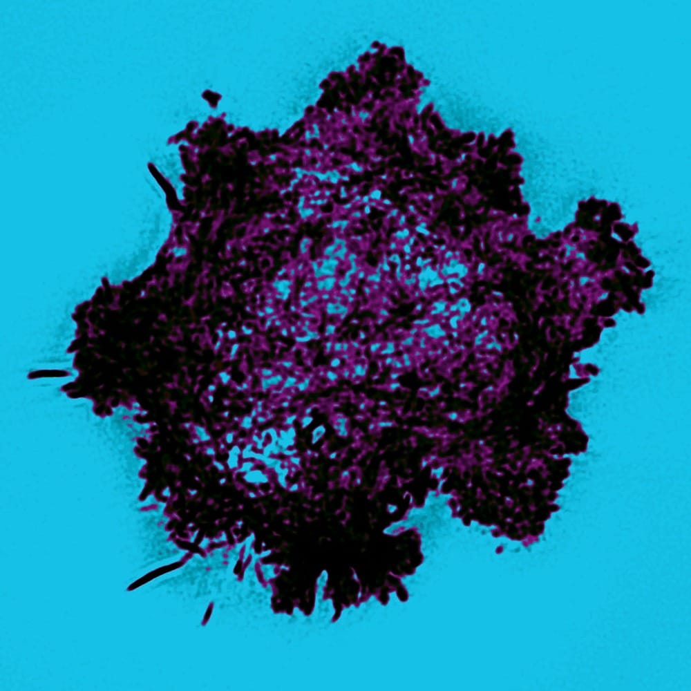
Cells captured in highest detail
Imperial and Oxford team up to tackle white blood cells

Researchers from Imperial College teamed up with the University of Oxford were able to reveal how white blood cells kill diseased tissue using deadly granules in more detail than ever before. The findings were published in the Public Library of Science (PLoS) and outline the use of optical laser tweezers and a microscope to be able to see the internals of white blood cells at the highest resolution ever.
The study involved white blood cells known as Natural Killers (NK) which play a major role in the rejection of tumours and infected cells. The research was led by Professor Daniel Davis from the Department of Life Sciences, who said that “NK cells may also play a role in the outcome of bone marrow transplants by determining whether a recipient’s body rejects or accepts the donated tissue.”
The results of this research will allow scientists to learn more about how NK cells identify which tissues to kill. “In the future, drugs that influence where and when NK cells kill could be included in medical treatments, such as the targeted killing of tumours”, Professor Davis added, “they may also prove useful in preventing the unwanted destruction by NK cells that may occur in transplant rejection or some autoimmune diseases.”
The research was conducted by using a novel imaging technique developed in collaboration with physicists at Imperial whilst using a super-resolution microscope at Oxford. An NK cell and its target were immobilised using a pair of laser tweezers so the microscope could capture the resulting action at the interface between the cells. Inside the NK cell, the actin filaments created a portal to which the enzyme filled granules moved towards, ready to pass through the NK cell in order to reach and kill the target. Dr Alice Brown of the Department of Life Sciences at Imperial said that “these previously undetectable events have never been seen in such high resolution.” She was one of the researchers who carried out many of the experiments involved, adding “it is truly exciting to observe what happens when an NK cell springs into action.”
The distance between an NK cell and its target when in contact is only about a hundredth of a millimetre across and the actin proteins and granules continuously change position until the target is killed. As a result, the super-resolution microscope had to be able to capture in such a frame rate and with such detail in order to successfully reveal their activity.
On the use of the microscope technologies developed with the Photonics Group, Professor Paul French from the Department of Physics at Imperial said “using laser tweezers to manipulate the interface between live cells into a horizontal orientation means our microscope can take many images of the cell contact interface in rapid succession”, providing an “unprecedented means to directly see dynamic molecular processes that go on between live cells”.
The method used maximises the amount of light captured from the cells allowing for super-resolution 3D images at twice the normal resolution of a conventional light microscope. Professor Ilan Davis, a Wellcome Trust Senior Fellow at the University of Oxford, involved in applying such techniques to cell biology research, said that their “microscope has given unprecedented views inside living NK cells”.
Funded by the Medical Research Council, the Biotechnology and Biological Sciences Research Council and a Marie Curie Intra-European Fellowship, the study also won a £150,000 award from the Rector’s Research Excellence Prize for high academic achievement in research with significant potential. Both Professors Daniel Davis and Paul French have Wolfson Royal Society Research Merit Awards.


